Effects of Malayan Green Dwarf Coconut Water on Platelets, White Blood Cells, and Liver Morphology in Iron Dextran-Induced Hemochromatosis in Wistar Rats
-
Emmanuel Chinedu Onuoha


Department of Haematology, Blood Transfusion Science, Faculty of Medical laboratory Science, Federal University, Otuoke, Bayelsa, Nigeria
Ezekiel Fayiah HallieSchool of Pharmacy, University of Liberia, Monrovia, Liberia
Oluebube Faith EzenwaforUniversity of South Carolina, Columbia, SC 29208, USA
| Received 05 Mar, 2025 |
Accepted 09 May, 2025 |
Published 10 May, 2025 |
Background and Objective: Iron overload, or hemochromatosis, is a pathological condition characterized by excessive iron deposition, leading to oxidative stress and organ damage. This study investigates the effects of Malayan Green Dwarf coconut water on hematological parameters and liver morphology in Wistar rats with iron dextran-induced hemochromatosis. Materials and Methods: Thirty Wistar rats were divided into 5 groups (n = 6 each): A negative control, a positive control (iron dextran-induced), and three treatment groups receiving 10, 20, and 30 mL/kg of Malayan Green Dwarf coconut water after iron overload induction. Hematological parameters (platelets, total white blood cells, and differentials) were analyzed, and liver tissues were examined histologically for morphological changes. Data were statistically analyzed using t-tests with significance set at p<0.05. Results: Iron overload resulted in reduced platelet count and lymphocytes while increasing white blood cell counts and inflammatory markers, though not statistically significant (p>0.05). Treatment with 10 mL/kg coconut water significantly reduced WBC count (p<0.05), while higher doses (20 and 30 mL/kg) preserved liver morphology by reducing iron deposition and hepatocellular damage. Conclusion: Malayan Green Dwarf coconut water exhibits potential hematoprotective and hepatoprotective effects in iron overload conditions. These findings suggest its role as a natural, cost-effective alternative for managing iron-induced toxicity, though further research is needed to confirm its mechanisms and clinical applicability.
| Copyright © 2025 Onuoha et al. This is an open-access article distributed under the Creative Commons Attribution License, which permits unrestricted use, distribution, and reproduction in any medium, provided the original work is properly cited. |
INTRODUCTION
Iron overload, a condition characterized by excessive iron accumulation in tissues, is a significant concern due to its potential to induce oxidative stress, inflammation, and tissue damage1. In both human and experimental models, excessive iron deposits have been linked to hepatic dysfunction, hematological abnormalities, and increased risk of fibrosis or cirrhosis2. The liver plays a crucial role in iron metabolism, making it highly susceptible to iron-induced toxicity, leading to hepatocellular damage and alterations in blood parameters3.
Iron dextran, commonly used in experimental models, mimics iron overload conditions seen in hereditary hemochromatosis, chronic transfusion therapy, and other iron-related disorders4. Excessive iron exposure promotes the production of free radicals via the Fenton reaction, which triggers oxidative stress and damages cellular components such as lipids, proteins, and DNA5. Consequently, iron overload can lead to significant changes in hematological parameters, including alterations in white blood cells, platelets, and differentials, which are indicative of immune responses and systemic inflammation6.
Natural compounds with antioxidant and hepatoprotective properties have gained attention for their potential in mitigating iron-induced toxicity. Among these, coconut water, particularly from the Malayan Green Dwarf variety, is rich in bioactive compounds such as cytokinin, L-arginine, and antioxidants that may offer protective effects against oxidative stress and inflammation7. Previous studies have demonstrated that coconut water possesses anti-inflammatory, detoxifying, and tissue-protective properties8. These characteristics suggest that coconut water may play a significant role in counteracting the hematological and hepatic disturbances caused by iron overload.
This study aims to evaluate the impact of Malayan Green Dwarf coconut water on platelet count, WBC differentials, and liver morphology in Wistar rats with iron dextran-induced hemochromatosis. By assessing hematological parameters and histological liver changes, the study seeks to determine whether coconut water administration can mitigate iron overload-related toxicities. The findings from this research could provide insights into alternative therapeutic strategies for managing iron overload disorders and related hepatic complications.
MATERIALS AND METHODS
Study area: This histology study was carried out at Medical Laboratory Science Department, Niger Delta University, Amassoma, while the complete blood count was carried out at Niger Delta University Teaching Hospital, Okolobiri both in Bayelsa State, Nigeria from January to June, 2024.
Study population: Thirty Wistar rats were bred in Animal House of the Department of Pharmacology, Niger Delta University, Amassoma, Bayelsa State were used in this study.
Ethical approval: College Health Research Ethics Committee, College of Health Sciences, Niger Delta University, Amassosoma, Bayelsa State, provided ethical clearance and approval.
Experimental design
Iron dextran solution: Iron dextran injection B.P injection 250 mg/5 mL, a product of Ancalima Lifesciences Ltd., Murthal, India, with batch number 1288010 was used in this research.
Acute toxicity study: The acute toxicity of Iron dextran was carried out using the standard method of Lorke9. The experiment was divided into two phases. In the phase one, 9 rats were divided into 3 groups of 3 rats each and were given 10, 100 and 1000 mg/kg of iron dextran intraperitoneally. In the phase two, 3 rats were divided into 3 groups of 1 rat each, and were given 1600, 2900, and 5000 mg/kg of iron dextran intraperitoneally after overnight fasting. For signs of toxicity such as paw-licking, stretching, respiratory stress, and mortality for the first 4 hrs, and the number of deaths per group were recorded after 24 hrs.
The LD50 was calculated using the formula:
Where:
| a | = | Least dose that caused mortality | |
| b | = | Highest dose that did not cause mortality |
Where:
| a | = | 1000 | |
| b | = | 100 | |
| = | 316.23 mg/kg |
Therefore, the median lethal dose (LD50) of acute toxicity test of iron dextran in Wistar rat was found to be 316.23 mg/kg b.wt.9.
Experimental design: This study used Thirty Wistar rats (170-200 g) bred in the animal house of the Department of Pharmacology, Niger Delta State, Welberforce Island, Bayelsa State. Before the experiment, the animals were allowed to be acclimatized in the animal house for approximately 7 days. They were housed in well-ventilated cages that were cleaned and food was replaced daily at a room temperature of about 37°C. The animals were fed with commercially prepared vital feed ad libitum and tap water. They were chosen at random and divided into 6 groups. Group 1 serves as the negative control, while Group 2 serves as the positive control. Group 1 received water and a normal diet. The groups 3, 4, and 5 were intraperitoneally administered 316.23 mg/kg b.wt., of iron dextran for 30 min, then 10, 20 and 30 mL/kg fresh coconut water were given via orogastric intubation, respectively.
| • | Group 1: Vital feed+water (negative control) | |
| • | Group 2: Induction with iron dextran+vital feed (positive control) | |
| • | Group 3: Induction with iron dextran+vital feed+coconut water (dose 1:10 mL/kg b.wt.) | |
| • | Group 4: Induction with iron dextran+vital feed+coconut water (dose 2:20 mL/kg b.wt.) | |
| • | Group 5: Induction with iron dextran+vital feed+coconut water (dose 3:30 mL/kg b.wt.) |
Collection of sample
Collection of blood samples: After 3 weeks of treatment, the rats were anesthetized by placing them in a glass chamber containing cotton wool soaked in chloroform, and they were then humanely sacrificed one by one. Blood samples (5 mL) were collected from the animals using cardiac puncture and dispensed into an EDTA bottle for platelets and white blood cells count. Platelets and white blood cells count were analyzed 8 hrs after sample collection.
Removal of organs: After the animals had been sacrificed, the livers were harvested and immediately fixed in 10% formalin saline solution for histological studies.
Determination of platelets and white blood cells count: Platelets and white blood cells count were determined by Automated Hematology Analyzer (SYSMEX XP-300).
Histological analysis of the liver: General cell morphology of the livers was determined using Haematoxylin and Eosin (H&E) staining techniques while iron pigment deposits were determined by the Perl’s Prussian blue staining technique.
Collection of coconut water: Fresh Malayan green dwarf Hybridized immature coconuts were harvested in the coconut farm in Opume, Ogbia local government of Bayelsa State based on the recent study10.
RESULTS
The comparison of platelets and white blood cells differentials between Wistar rat without iron dextran Induction (negative control) group and iron dextran induced haemochromatosis in Wistar rat (positive control) group as shown in Table 1. Platelet count and lymphocytes were lower in the iron dextran-induced haemochromatosis Wistar rat (positive control) group than in the negative control group, but they were not statistically significant (p>0.05). However, the total white blood cells count, neutrophils, monocytes, and eosinophils were higher in iron dextran induced haemochromatosis Wistar rat (positive control) group than the negative control group, but not statistically significant (p>0.05).
Table 2 shows the comparison of platelets and white blood cells differentials between iron dextran induced haemochromatosis in Wistar rat (positive control) group and treatment group with administration of 10 mL/kg b.wt., of Malayan green dwarf hybridized immature coconut water after induction of iron dextran in Wistar rat (positive control). The total white blood cells counts (3.37±2.63) were significantly lower in the treatment group (10 mL/kg) than iron dextran induced haemochromatosis Wistar rat (positive control) (14.0±2.00) group (p<0.05) while platelet count, lymphocytes, neutrophils, monocytes and eosinophils were not significant (p>0.05).
The comparison of platelets and white blood cells differentials between iron dextran induced haemochromatosis in Wistar rat (positive control) group and treatment group with administration of 20 mL/kg b.wt., of Malayan green dwarf hybridized immature coconut water after induction of iron dextran in Wistar rat (positive control) as shown in Table 3. Platelet count, total white blood cells, and lymphocytes were lower in iron dextran induced haemochromatosis Wistar rat (positive control) group than the treatment group (20 mL/kg) but not statistically significant (p>0.05). However, Neutrophils, Monocytes, and Eosinophils were higher in iron dextran-induced haemochromatosis Wistar rat (positive control) group than treatment group (20 mL/kg) but not statistically significant (p>0.05).
| Table 1: | Comparison of platelets, wbcs, and wbc differentials in wistar rats with and without iron dextran-induced haemochromatosis (N = 6) | |||
| Haematology parameter | Negative control | Positive control | t-value | p-value |
| Platelets (109/L) | 872.7±66.5 | 620.0±300.9 | 1.85 | 0.206 |
| White blood cells (109/L) | 8.83±2.7 | 14.0 ±2.00 | -2.14 | 0.166 |
| Lymphocytes (%) | 77.00±2.0 | 71.00±7.810 | 1.27 | 0.332 |
| Neutrophils (%) | 12.33±0.58 | 14.00±3.464 | -0.71 | 0.549 |
| Monocytes (%) | 7.33±1.53 | 11.00±3.464 | -1.808 | 0.212 |
| Eosinophil (%) | 3.33±0.58 | 4.00±1.000 | -1.000 | 0.423 |
| Significant differences at p<0.05 level | ||||
| Table 2: | Blood cell comparison between haemochromatosis and treatment groups (10 mL/kg) in Wistar rats (N = 6) | |||
| Haematology parameter | Treatment (10 mL/kg) | Positive control | t-value | p-value |
| Platelets (109/L) | 691.33±226.6 | 620.0±300.9 | -0.289 | 0.8 |
| White blood cells (109/L) | 3.37±2.63* | 14.0±2.00* | 6.901 | 0.02 |
| Lymphocytes (%) | 73.00±6.56 | 71.00±7.810 | -0.378 | 0.742 |
| Neutrophils (%) | 18.33±5.51 | 14.00±3.46 | -1.182 | 0.359 |
| Monocytes (%) | 6.67±1.15 | 11.00±3.46 | 2.46 | 0.133 |
| Eosinophil (%) | 2.00±0.00 | 4.00±1.00 | 3.46 | 0.074 |
| *Significant differences at p<0.05 level | ||||
|
| Table 3: | Blood cell comparison between haemochromatosis and treatment groups (20 mL/kg) in Wistar rats (N = 6) | |||
| Haematology parameter | Treatment (20 mL/kg) | Positive control | t-value | p-value |
| Platelets (109/L) | 698.67±129.27 | 620.0±300.9 | -0.794 | 0.511 |
| White blood cells (109/L) | 15.43±0.96 | 14.0±2.00 | -1.323 | 0.317 |
| Lymphocytes (%) | 79.67±6.51 | 71.00±7.81 | -1.562 | 0.259 |
| Neutrophils (%) | 11.67±3.51 | 14.00±3.46 | 0.788 | 0.513 |
| Monocytes (%) | 6.00±2.00 | 11.00±3.46 | 2.17 | 0.163 |
| Eosinophil (%) | 2.67±1.16 | 4.00±1.00 | 4.00 | 0.057 |
| Significant differences at p<0.05 level | ||||
| Table 4: | Blood cell comparison between haemochromatosis and treatment groups (30 mL/kg) in Wistar rats (N = 6) | |||
| Haematology parameters | Treatment (30 mL/kg) | Positive control | t-value | p-value |
| Platelets (109/L) | 865.33±108.28 | 620.0±300.9 | 1.56 | 0.26 |
| White blood cells (109/L) | 10.80±3.25 | 14.0±2.00 | 1.06 | 0.402 |
| Lymphocytes (%) | 75.33±3.055 | 71.00±7.81 | -0.702 | 0.555 |
| Neutrophils (%) | 13.00±1.00 | 14.00±3.464 | 0.655 | 0.58 |
| Monocytes (%) | 8.67±2.891 | 1.00±3.46 | 0.636 | 0.59 |
| Eosinophil (%) | 3.00±1.00 | 4.00±1.00 | 1.000 | 0.423 |
| Significant differences at p<0.05 level | ||||
Table 4 shows the comparison of platelets, and white blood cells differentials between Wistar rat without iron dextran Induction (negative control) group and iron dextran induced haemochromatosis in Wistar rat (positive control) group. Platelet count and lymphocytes were lower in iron dextran induced haemochromatosis Wistar rat (positive control) group than treatment group (30 mL/kg) but not statistically significant (p>0.05). However, the total white blood cells count, neutrophils, monocytes, and eosinophils were higher in iron dextran-induced haemochromatosis Wistar rat (positive control) group than the treatment group (30 mL/kg) but not statistically significant (p>0.05).
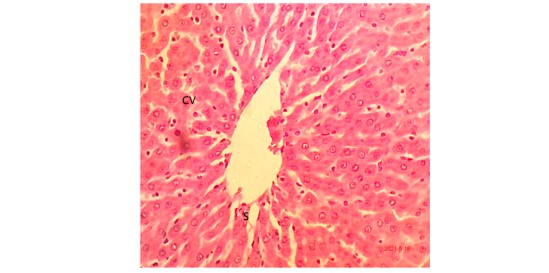
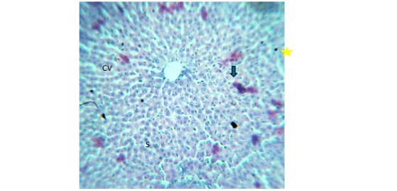
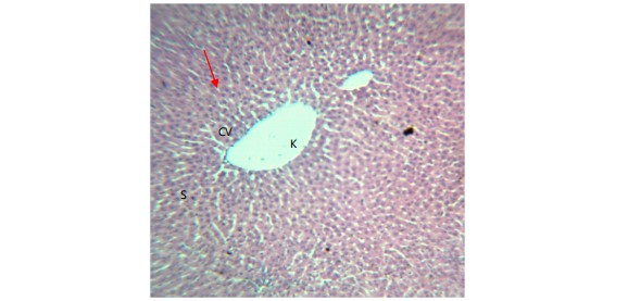
The morphology of liver tissues under different conditions is illustrated. Figure 1 depicts the negative control (vital feed+water), showing normal hepatic architecture with hepatocytes arranged in cords radiating from the centra vein (CV) and arrow, intact sinusoidal spaces (S). Hepatocytes (H) are polyhedral in shape (×10) H&E. Figure 2 represents the positive control (iron dextran-induced without coconut water treatment), revealing binucleated hepatocytes w ith granular iron deposits in Kupffer cells (yellow star) and congestion of the portal vein.
|
|
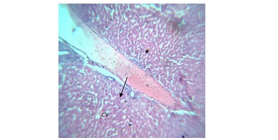
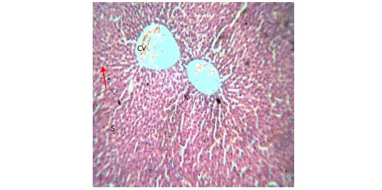
Figure 3 illustrates iron dextran-induced liver following administration of 10 mL/kg b.wt., of tender coconut water, showing granular degeneration of hepatocytes, congestion of the portal vein, and the presence of Kupffer cells. Figure 4 presents the liver after iron dextran induction and administration of vital feed with coconut water (Dose 2: 20 mL/kg b.wt.), exhibiting mild distortion of the central vein (CV) walls (red arrow), slightly congested central vein, mild granular degeneration of hepatocytes, intact sinusoidal spaces (S), and the presence of Kupffer cells. Figure 5 displays the liver after iron dextran induction and administration of Vital feed with coconut water (Dose 3: 30 mL/kg b.wt.), showing mild distortion of the central vein, a clear central vein, intact sinusoidal spaces (S), and the presence of Kupffer cells.
|
|
DISCUSSION
Iron overload has been widely associated with hematological alterations, including thrombocytopenia, leukocytosis, and changes in differential WBC counts2. The present study found that platelet count and lymphocytes were lower in iron dextran-induced hemochromatosis Wistar rats compared to the negative control, though not statistically significant (p>0.05). This is consistent with findings in which excessive iron accumulation suppresses thrombopoiesis due to oxidative stress-induced bone marrow suppression11.
Additionally, increased WBC count, neutrophils, monocytes, and eosinophils observed in the iron-overloaded rats support previous studies that link iron overload to inflammation and immune system activation12. Elevated monocytes and neutrophils are indicative of oxidative stress and inflammatory responses triggered by excessive iron deposits in tissues13.
Following treatment with Malayan Green Dwarf coconut water, a significant reduction (p<0.05) in WBC count was observed at 10 mL/kg, suggesting its potential anti-inflammatory effects. This is supported by a study that demonstrated that coconut water contains bioactive compounds such as flavonoids and vitamin C, which have antioxidant and anti-inflammatory properties14. However, at higher doses (20 and 30 mL/kg), the hematological changes were not statistically significant, suggesting a possible saturation point for the therapeutic effect of coconut water.
Histological analysis of liver tissues revealed marked iron deposition, binucleated hepatocytes, and congestion of the portal vein in iron-overloaded rats. This aligns with previous studies showing that iron accumulation in the liver causes hepatocellular damage, fibrosis, and Kupffer cell activation1. Excess iron is known to catalyze the formation of reactive oxygen species (ROS), leading to oxidative stress, lipid peroxidation, and hepatic inflammation15.
Administration of coconut water showed a dose-dependent improvement in liver morphology. At 10 mL/kg, signs of granular degeneration of hepatocytes and congestion of the portal vein were still evident, indicating mild hepatotoxicity. However, at 20 and 30 mL/kg, the liver showed reduced iron deposition, clearer sinusoidal spaces, and less severe morphological alterations. These findings are in agreement with studies by researchers who reported that coconut water has hepatoprotective properties due to its high potassium, amino acid, and antioxidant content16,17.
The protective effect of coconut water observed in this study can be attributed to its ability to mitigate oxidative stress and inflammation. Studies have shown that polyphenols and flavonoids present in coconut water act as natural iron chelators, helping to reduce free iron levels and prevent oxidative damage18. Additionally, the electrolyte composition of coconut water helps maintain cellular homeostasis, which is crucial in counteracting the toxic effects of iron overload3.
CONCLUSION
The findings of this study demonstrate that Malayan Green Dwarf coconut water has potential hematoprotective and hepatoprotective effects in iron dextran-induced hemochromatosis in Wistar rats. This study confirms that iron overload is associated with hematological alterations, including increased WBC count, neutrophils, monocytes, and eosinophils, indicative of inflammation and oxidative stress. Platelet count and lymphocytes were lower in iron-overloaded rats, though not statistically significant. Histological analysis revealed severe hepatic iron deposition and structural damage. Treatment with Malayan Green Dwarf coconut water at 10 mL/kg significantly reduced WBC count (p<0.05), suggesting anti-inflammatory effects, while higher doses showed diminished impact, indicating a saturation point. Liver morphology improved in a dose-dependent manner, with reduced iron deposition at 20 and 30 mL/kg, supporting its hepatoprotective role. Future research should explore the long-term efficacy of coconut water in managing iron overload and its potential in clinical applications. Further investigations are needed also to fully elucidate the biochemical mechanisms and to explore potential clinical applications.
SIGNIFICANCE STATEMENT
The significance of this study lies in its demonstration of the potential benefits of Malayan Green Dwarf coconut water in mitigating hematological and hepatic complications of iron overload. By providing evidence of its antioxidant, anti-inflammatory, a nd hepatoprotective properties, this research contributes to the growing body of knowledge on natural remedies for iron-induced toxicity. It opens new avenues for developing cost-effective and accessible treatments for managing hemochromatosis and related conditions.
REFERENCES
- Papanikolaou, G. and K. Pantopoulos, 2005. Iron metabolism and toxicity. Toxicol. Applied Pharmacol., 202: 199-211.
- Brissot, P., M.B. Troadec, O. Loréal and E. Brissot, 2019. Pathophysiology and classification of iron overload diseases; update 2018. Transfus. Clin. Biol., 26: 80-88.
- Ganz, T., 2013. Systemic iron homeostasis. Physiol. Rev., 93: 1721-1741.
- Gozzelino, R. and P. Arosio, 2016. Iron homeostasis in health and disease. Int. J. Mol. Sci., 17.
- Zaric, B.L., M.T. Macvanin and E.R. Isenovic, 2023. Free radicals: Relationship to human diseases and potential therapeutic applications. Int. J. Biochem. Cell Biol., 154.
- Kouroumalis, E., I. Tsomidis and A. Voumvouraki, 2023. Iron as a therapeutic target in chronic liver disease. World J. Gastroenterol., 29: 616-655.
- Tan, T.C., L.H. Cheng, R. Bhat, G. Rusul and A.M. Easa, 2014. Composition, physicochemical properties and thermal inactivation kinetics of polyphenol oxidase and peroxidase from coconut (Cocos nucifera) water obtained from immature, mature and overly-mature coconut. Food Chem., 142: 121-128.
- Manna, K., A. Khan, D.K. Das, S.B. Kesh and U. Das et al., 2014. Protective effect of coconut water concentrate and its active component shikimic acid against hydroperoxide mediated oxidative stress through suppression of NF-κB and activation of Nrf2 pathway. J. Ethnopharmacol., 155: 132-146.
- Lorke, D., 1983. A new approach to practical acute toxicity testing. Arch. Toxicol., 54: 275-287.
- Onuoha, E.C. and E.F. Hallie, 2024. Comparative study of different species of coconut water and their health benefits. Int. J. Innovative Appl. Res., 12: 30-37.
- Xie, X., L. Chang, X. Zhu, F. Gong and L. Che et al., 2025. Rubiadin mediates the upregulation of hepatic hepcidin and alleviates iron overload via BMP6/SMAD1/5/9-signaling pathway. Int. J. Mol. Sci., 26.
- di Paola, A., C. Tortora, M. Argenziano, M.M. Marrapodi and F. Rossi, 2022. Emerging roles of the iron chelators in inflammation. Int. J. Mol. Sci., 23.
- Visitchanakun, P., W. Panpetch, W. Saisorn, P. Chatthanathon and D.L. Wannigama et al., 2021. Increased susceptibility to dextran sulfate-induced mucositis of iron-overload β-thalassemia mice, another endogenous cause of septicemia in thalassemia. Clin. Sci., 135: 1467-1486.
- DebMandal, M. and S. Mandal, 2011. Coconut (Cocos nucifera L.: Arecaceae): In health promotion and disease prevention. Asian Pac. J. Trop. Med., 4: 241-247.
- Raffaeli, G., F. Manzoni, V. Cortesi, G. Cavallaro, F. Mosca and S. Ghirardello, 2020. Iron homeostasis disruption and oxidative stress in preterm newborns. Nutrients, 12.
- Parmar, P.T., A.K. Singh and S.G. Borad, 2021. Coconut (Cocos nucifera). In: Oilseeds: Health Attributes and Food Applications, Tanwar, B. and A. Goyal (Eds.), Springer, Singapore, ISBN: 978-981-15-4194-0, pp: 163-189.
- Kontoghiorghes, G.J. and C.N. Kontoghiorghe, 2020. Iron and chelation in biochemistry and medicine: New approaches to controlling iron metabolism and treating related diseases. Cells, 9.
- O’Brien, B.J., L.R. Bell, D. Hennessy, J. Denham and C.D. Paton, 2023. Coconut water: A sports drink alternative? Sports, 11.
How to Cite this paper?
APA-7 Style
Onuoha,
E.C., Hallie,
E.F., Ezenwafor,
O.F. (2025). Effects of Malayan Green Dwarf Coconut Water on Platelets, White Blood Cells, and Liver Morphology in Iron Dextran-Induced Hemochromatosis in Wistar Rats. Pharmacologia, 16(1), 5-13. https://doi.org/10.17311/pharma.2025.05.13
ACS Style
Onuoha,
E.C.; Hallie,
E.F.; Ezenwafor,
O.F. Effects of Malayan Green Dwarf Coconut Water on Platelets, White Blood Cells, and Liver Morphology in Iron Dextran-Induced Hemochromatosis in Wistar Rats. Pharmacologia 2025, 16, 5-13. https://doi.org/10.17311/pharma.2025.05.13
AMA Style
Onuoha
EC, Hallie
EF, Ezenwafor
OF. Effects of Malayan Green Dwarf Coconut Water on Platelets, White Blood Cells, and Liver Morphology in Iron Dextran-Induced Hemochromatosis in Wistar Rats. Pharmacologia. 2025; 16(1): 5-13. https://doi.org/10.17311/pharma.2025.05.13
Chicago/Turabian Style
Onuoha, Emmanuel, Chinedu, Ezekiel Fayiah Hallie, and Oluebube Faith Ezenwafor.
2025. "Effects of Malayan Green Dwarf Coconut Water on Platelets, White Blood Cells, and Liver Morphology in Iron Dextran-Induced Hemochromatosis in Wistar Rats" Pharmacologia 16, no. 1: 5-13. https://doi.org/10.17311/pharma.2025.05.13

This work is licensed under a Creative Commons Attribution 4.0 International License.