Biochemical Effects of Nauclea latifolia Ethanolic Root Extract in Rats
-
M.C. Udeh Sylvester
Department of Biochemistry, Federal University Wukari, 640102, Wukari, Taraba, Nigeria
V.N. OguguaDepartment of Biochemistry, University of Nigeria, 410105, Nsukka, Enugu, Nigeria
E.Y. Ojochenemi
Department of Biochemistry, Federal University Wukari, 640102, Wukari, Taraba, Nigeria
E.J. Parker
Department of Biochemistry, University of Nigeria, 410105, Nsukka, Enugu, Nigeria
S. EgbaDepartment of Biochemistry, Federal University Wukari, 640102, Wukari, Taraba, Nigeria
E. AnaduakaDepartment of Biochemistry, University of Nigeria, 410105, Nsukka, Enugu, Nigeria
O.P. UgwuDepartment of Biochemistry, University of Nigeria, 410105, Nsukka, Enugu, Nigeria
Michael Sunday Abu 
Department of Biochemistry, Federal University Wukari, 640102, Wukari, Taraba, Nigeria
T.J. IornengeDepartment of Biochemistry, Federal University Wukari, 640102, Wukari, Taraba, Nigeria
| Received 29 Jul, 2022 |
Accepted 21 Dec, 2022 |
Published 31 Mar, 2023 |
Background and Objective: The plant Nauclea latifolia (Smith) (Family: Rubiaceae) also known as ‘Pin Cushion tree’ or ‘African Peach’ is a struggling shrub, native to tropical Africa and Asia where the use of folk medicine is prevalent and the search for a herbal cure is but common practice. This report described the biochemical effects of Nauclea latifolia ethanolic root extract in rats. Materials and Methods: Extraction of the root of Nauclea latifolia with ethanol was carried out. A total of twenty Wister albino rats were used in this study. The rats were randomly divided into four groups of five rats each. The various biochemical tests were then conducted on the experimental rats. Results: The acute toxicity study on ethanol extract in mice established an intraperitoneal LD50 greater than 4000 mg kg–1. The phytochemical analysis of the Nauclea latifolia ethanol extract indicated the presence of flavonoids, saponins and reducing sugars. The liver alkaline phosphatase, alanine aminotransferase, aspartate aminotransferase and the serum concentrations of conjugated and total bilirubin measured suggest mild damage to the liver hepatocytes of rats treated with ethanolic extract of Nauclea latifolia root when compared to the control. The result of urea and creatinine indicate that the kidney may be damaged when compared to the control group. The extract caused a slight but non-significant increase (p>0.05) in blood glucose concentration as administration of the extract continued on days 28 and 42. Conclusion: The present study established that the continuous administration of the ethanol crude extract ofNauclea latifolia is lethal to the hepatocytes and kidneys.
| Copyright © 2023 Udeh Sylvester et al. This is an open-access article distributed under the Creative Commons Attribution License, which permits unrestricted use, distribution, and reproduction in any medium, provided the original work is properly cited. |
INTRODUCTION
The large quantities of minerals, primary and secondary metabolites and several claimed therapeutic characteristics of medicinal plants make them useful to humanity1. The discovery of phytochemicals in traditional medicinal plants offers a promising possibility for the creation of novel therapies. Ageing, oxidative stress and the onset of chronic illnesses can all be significantly impacted by phytochemicals. Numerous physical and metabolic processes are bio-enhanced by phytoprotectants2.
The multi-stemmed Nauclea latifolia tree may reach a height of 200 m. In Nigeria, it is marketed under the trade name Opepe and is also referred to as the Pin Cushion tree and the African peach in English3. Locally, it is referred to as Itu by the Itsekiris in Edo State and Mbomibong by the Ibibios and Effik in Akwa-Ibom State in the South-South of Nigeria. It is referred to as Tafashiya or Tafiyayaiga in Hausa, Northern Nigeria and Uche by the Igedes in Benue State. It is referred to as Ubulinu or Ovoroilu in Ibo, South Eastern Nigeria and Egbeyesi or Egbesi in Yoruba, South West Nigeria4.
Diabetes and hypertension treatments are two instances of ethnomedical4. Cold infusion of the stem bark is used as an anthelmintic and diuretic in Northern Nigeria. It is utilised as a chewing stick, mouthwash and treatment for TB and gastrointestinal3. There have been reports of its use as an aphrodisiac, vermifuge, antimalarial and antipyretic5. Almost every component of the plant may be used to cure diseases. In ethnomedicine, the order of use is roots>stem>bark>leaf4. The anti-malaria6, antipyretic, antiinflammatory and antinociceptive7, anthelminthic8,9, wound healing and antitrypanosomal5 of Nauclea latifolia have been evaluated. This report was therefore focused on the biochemical effects of Nauclea latifolia ethanolic root extracts in rats.
MATERIALS AND METHODS
Study area: The study was carried out at the Biochemistry Department, University of Nigeria, Nsuka, Enugu State, Nigeria from October, 2019-2020.
Animal material: The experimental animals used for the study were male and female Wister albino rats (weights 103-230 g). The rats were obtained from the animal house of the Faculty of Veterinary Medicine, University of Nigeria, Nsukka and were kept for 7 days to acclimatize and given feed and water ad libitum.
Plant material: Nauclea latifolia roots were obtained from a forest in Okpome-Agbada Nenwe, Aninri Local Government Area of Enugu State, Nigeria. The roots were authenticated by Mr. Njokuocha in the Department of Botany, University of Nigeria, Nsukka. The roots were dried at room temperature in the absence of sunlight for 1 week, cut into bits and ground to a coarse powder using a hammer mill.
Extraction procedure
Preparation of ethanolic extract: About 500 g of the powdered root of Nauclea latifolia was soaked in 3.4 L of 70% ethanol. The mixture was left to stand for 24 hrs with occasional stirring. The mixture was later extracted using a soxhlet extractor to obtain the ethanol extract. The extract was concentrated over a water bath at a temperature of 25 to 30°C to obtain 46 g (9.2%) of the ethanol extract.
Determination of concentration of extract: A crucible was weighed and a known weight of the extract was poured into the crucible and their combined weight was determined before heating. The crucible was heated until all its content was charred. After heating, the crucible was re-weighed with its content and the weight was recorded. The concentration of the solid extract was then determined from the two weights.
Experimental design: A total of sixty Wister albino rats were used in this study. The rats were randomly divided into four groups of 5 rats each.
Group 1 : |
Control group was administered normal saline |
Group 2 : |
Administered 10 mg kg–1 of the ethanolic extract |
Group 3 : |
Administered 50 mg kg–1 of the ethanolic extract |
Group 4 : |
Administered 250 mg kg–1 of the ethanolic extract |
The extracts were administered by dissolving accurately measured doses in water and feeding the rats orally daily for 42 days. In the end, the rats were sacrificed and some internal organs were removed, fixed and later used for histopathological studies.
Acute toxicity test (LD50): The Median Lethal Dose (LD50) of the ethanol extract was determined in rats using the oral route of administration10. A total of thirty albino Wister rats were randomly divided into six groups of five rats. Each rat was kept in a separate clean stainless cage and provided with water and feed ad libitum. The rats in the various groups were injected intraperitoneally with increasing doses of 250, 500, 1000, 2000 and 4000 mg kg–1 body weight of the Nauclea latifolia ethanolic extract while the control group received an equal volume of distilled water. The rats were allowed to feed and were closely observed for mortality and toxic signs for 5 days. No death was recorded. Since the extract did not cause any death in the rats at concentrations above 4000 mg kg–1, the ethanolic extract of Nauclea latifolia was considered non-toxic10.
Cytotoxicity test (LC50): Artemia salina eggs obtained from Dr. A.O. Onaga, of the Department of Veterinary Pharmacology and Physiology, were incubated in natural seawater (from Bar Beach, Lagos Nigeria) in a dam-well under room condition. AS 48 hrs nauplii (10 in each well) were incubated in known concentrations of 10, 100 and 1000 ppm of the extract for 24 hrs. All tests were conducted in triplicate. The dead nauplii were subtracted from 10 and the data were analysed using profit analysis (Finner Program, DOS) to determine the LC50 at a 95% confidence interval. Weak nauplii were noted as an indication of central nervous system depressions while the control was incubated in seawater, the incubation medium.
Phytochemical analysis: The preliminary analysis involved testing for the presence or absence of the following plant constituents: Alkaloids, flavonoids, reducing sugars, saponins, tannins, acidity, oils and terpenoids using the method of Harborne11.
Test for the presence of alkaloids: Exactly 0.2 g of the ground dried roots of Nauclea latifolia were boiled with 5 mL of 2% hydrochloric acid in the steam bath. The mixture was filtered using Whatman No.1 filter paper. One millilitre of the solution was treated with two drops of Drahendroff reagent (Bismuth potassium iodide solution). A red precipitate indicated the presence of alkaloids11.
Test for the presence of reducing sugars: Exactly 0.1 g of the ground dried roots of Nauclea latifolia were shaken vigorously in 5 mL of distilled water and filtered. One mL portion of the filtrate was added to equal volumes of Fehling’s solutions A and B and then shaken vigorously. A brick red precipitate indicated the presence of reducing sugars.
Test for the presence of flavonoids: Approximately 0.2 g of the ground dried sample was heated with 10 mL of ethyl acetate in boiling water for 3 min. The mixture was filtered and the filtrate was used for the test. Four millilitres of the filtrate were shaken with 1 mL of 1% aluminium chloride solution and observed. Yellow colouration in the ethyl acetate layer indicated the presence of flavonoids.
Test for the presence of tannins: Exactly 2 g of the ground material was boiled in 5 mL of 45% ethanol for 5 min. The mixture was cooled and filtered. To 1 mL of the filtrate was added 3 drops of lead sub-acetate solution. A gelatinous precipitate indicated the presence of tannins.
Test for the presence of saponins: The ground sample (0.1 g) was boiled in 6 mL of distilled water for 5 min. The mixture was filtered while still hot. The filtrate was then used for the following tests:
Emulsion test: To 1 mL of the filtrate was added two drops of olive oil and shaken. The formation of emulsion indicated the presence of saponins
Frothing test: To 1 mL of the filtrate was mixed with 4 mL of distilled water and then shaken vigorously. Observation of stable frost on standing showed the presence of saponins
Tests for acidity: Extract (0.1 g) was placed in a clear dry test tube and sufficient distilled water was added. This was warmed in a water bath and then cooled. A strip of water-wetted litmus paper was dipped into the filtrate. The red colour on the litmus paper indicated acidity.
Tests for oils: The plant extract (0.1 g) was dropped on a filter paper and kept to dry. The filter paper was then observed for the presence of oils.
Tests for terpenoids: The ethanol extract (0.5 mL) was evaporated to dryness in a water bath and heated with 3 mL of concentrated sulphoric acid for 10 min in a water bath. A grey colour was a positive indicator.
Biochemical assay methods
Glucose concentration: A one-touch glucometer (Lifescan, USA) and test strips were used for this assay. The principle of the reaction is based on the glucose oxidase reaction. The blood glucose estimation involves the reaction between glucose in the blood and oxygen in the presence of glucose oxidase, which is immobilised in the test strip, to yield gluconic acid and hydrogen peroxide. The hydrogen peroxide subsequently oxidizes the dye in a reaction mediated by peroxidase to produce a blue-coloured product. The intensity of the colour produced is proportional to the glucose concentration in the blood. The colour intensity was read instantly from the one-touch glucometer.
Determination of Alkaline Phosphatase (ALP): The principle of this method is based on the reaction between serum alkaline phosphatase and a colourless substrate of phenolphthalein monophosphate, giving rise to phosphoric acid and phenolphthalein which at alkaline pH values, turn to pink that can be determined photometrically. The blank and sample test tubes were set up in duplicates. Into the sample test tubes, 0.5 mL distilled water and 0.01 mL substrate were pipetted, respectively and mixed thoroughly. Into the blank tube, 0.5mL distilled water and 0.01 mL of substrate were incubated at 37°C for 5 min. From the standard test tube, 0.1 mL was withdrawn and used as a blank. From the sample tube, 0.05 mL was withdrawn and incubated at 37°C for 20 min. The absorbance of the sample and the standard were measured against the blank at 550 nm.
ALP calculation: Alkaline phosphatase was calculated as follows:
Determination of Alanine Aminotransferase (ALT): The blank and sample test tubes were set up in duplicates. Into the blank sample tubes, 0.1 mL distilled water and 0.5 mL serum were pipetted, respectively. To these was added 0.5 mL solution from one of the kits containing phosphate buffer, L-alanine and ά-oxoglutarate at the concentrations specified above. The mixtures were thoroughly mixed and incubated for exactly 30 min at 37°C. Five millilitres of the solution containing 2, 4-dinitrophenylhydrazine (0.2 mmol L–1) was added to each tube, mixed thoroughly and incubated for exactly 20 min at 25°C. Sodium hydroxide (2.5 mL) was then added and the solution was mixed and the absorbance of the sample read against the blank after 5 min. The activity of ALT (U L–1) was read up from the standard reference table.
Determination of Aspartate Aminotransferase (AST): The blank and sample test tubes were set up in duplicates. Into the sample tube was pipetted 0.1 mL of sample and 0.5 mL of solution-1. Into the blank tube was pipetted 0.5 mL of solution-1 which was then mixed thoroughly and incubated for 30 min at 37°C. Then 0.5 mL of solution-2 and 0.1 mL of the sample were pipetted into a blank test tube. Also, 0.5 mL of solution-2 was pipetted into a sample tube, mixed thoroughly and allowed to stand for 20 min at 25°C. Twenty-five millilitres of sodium hydroxide was added into each test tube and mixed thoroughly. The absorbance was then read at 550 nm against the blank after 5 min. The activity of AST (U L–1) was read up from the standard reference table.
Determination of Total Bilirubin (TB): The blank and sample test tubes were set in duplicates. Into the blank sample tube, 0.2 mL of reagent-1, one millilitre of reagent-3 and 0.2 mL of sample were pipetted. Into the sample test tube, 0.2 mL of reagent-1, 0.05 mL of reagent-2, 1 mL of reagent-3 and 0.2 mL of sample were pipetted. The different tubes were mixed thoroughly and allowed to stand for 10 min at 25°C. Then 1 mL of reagent-4 was added to each of the test tubes. The tubes were allowed to stand for 10 min at 25°C and the absorbance read at 578 nm against the blank.
TB calculation:
where, ATB is Absorbance of total bilirubin 185 and 10.8 are constants.
Determination of Conjugated Bilirubin (CB): The blank and sample test tubes were set up in duplicates. Into the sample blank test tube, 0.2 mL of reagent-1, 2 mL of sodium chloride (9 g dL–1) and 0.2 mL of sample were added. Into the sample test tube, 0.2 mL of reagent-1, 0.05 mL of reagent-2, 2 mL of sodium chloride (9 g dL–1) and 0.2 mL of sample were added. The contents of the test tubes were mixed thoroughly and allowed to stand for 5 min at 20°C and the absorbance read at 546 nm against the blank.
CB calculation:
where, ACB is Absorbance of conjugated bilirubin 246 and 14.4 are constants.
Determination of serum urea: Three test tubes were labelled blank, standard and sample respectively and the solutions were added. The tubes were set up in duplicates and the contents were mixed thoroughly and incubated for 10 min at 37°C. Solution-2 and distilled water (25 mL) were, respectively added to each tube and the contents were shaken vigorously and incubated for 15 min at 37°C. The absorbance of each tube was read against the blank at 510 nm.
Calculation: The urea concentration in the serum was calculated using the formula:
Determination of serum creatinine: Exactly 0.1 mL of the sample was pipetted into a clean, labelled tube and 1.0 mL of trichloroacetic acid (TCA) was added to it, mixed and then centrifuged at 250 rpm for
10 min. The supernatant was decanted and reserved for use. By this procedure, the sample had been deproteinised. The assay procedure was then carried out.
The mixtures were prepared in duplicates, thoroughly mixed and allowed to stand for 20 min. The absorbance of the samples and standard were read against the blank at 540 nm.
Calculation: The concentration of creatinine in serum was calculated as follows:
RESULTS
Extraction: The ethanolic extract weighed 46 g, thus giving a 9.2% yield from the 500 g raw plant material used.
Phytochemical analysis: The qualitative phytochemical tests carried out on the ethanolic extract of N. latifolia showed the presence of the following compounds in Table 1. Reducing sugar was present in very high concentrations (+++) while flavonoids and terpenoids were resent in moderately high concentrations (++).
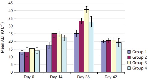
Effects of Nauclea latifolia ethanolic root extract on biiochemical parameters: The effect of the extract on the ALT was shown in Fig. 1. On days 0 and 42, the ethanolic extract of N. latifolia had no significant effect (p>0.05) on the concentration of ALT in rats compared to the control. On day 14 and 28, ALT activity was observed more in group-2 (10 mg kg–1) and group-3 (50 mg kg–1) showing a significant increase (p<0.05) in the concentration of ALT compared to the control.
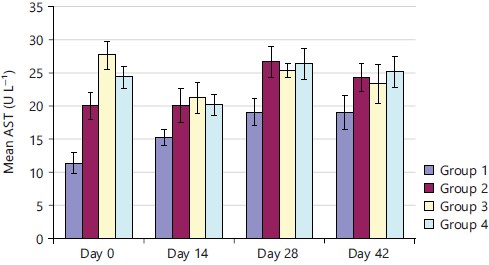
The administration of the ethanolic extract of N. latifolia caused a significant increase (p<0.05) in AST concentrations on all the treatment days compared to the control group as shown in Fig. 2. There was a remarkable dose-dependent increase in AST activity from day 14 to day 42. This implied that the administration of ethanolic extract had an increased effect on AST concentration by doses and by the number of days.
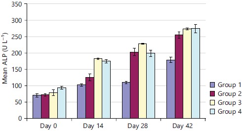
In Fig. 3, the ethanol extract did not affect the concentration of ALP (p>0.05) on day 0 compared to the control but on days 14, 28 and 42, the extract caused a significant increase (p<0.05) in the concentration of ALP compared to the control implying that continuous administration of the ethanol extract caused an increase in the ALP concentration.
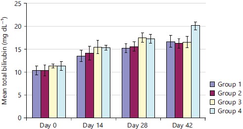
As demonstrated in Fig. 4, on day 0, the total bilirubin levels showed no significant difference (p>0.05) in groups 2, 3 and 4. There were slight increases in the level of total bilirubin among all groups from days 14, 28 to 42 when compared to the control group while group-4 (250 mg kg–1) on day 42 had a significantly higher concentration of total bilirubin compared to other groups.
| Table 1: | Phytochemical constituents of Nauclea latifolia ethanolic root extract | |||
| Alkaloids | - |
| Reducing sugars | +++ |
| Flavonoids | ++ |
| Tannins | - |
| Saponins | + |
| Acidity | - |
| Fats and oils | - |
| Terpenoids | ++ |
| +: >Present in trace concentration, ++: >Present in moderately high concentration, +++: >Present in very high concentration and -: >Absent | |
|
|
|
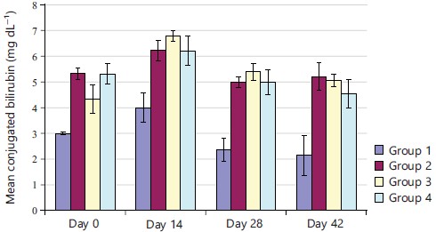
Conjugated bilirubin levels of rats treated with ethanolic extract were significantly higher (p<0.05) than those of the control rats across all the days in Fig. 5. The highest concentrations were observed on day 14 with group 3 showing the highest concentration. There was a gradual decline from day 14 in conjugated bilirubin concentration among the four groups.
|
|
|
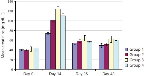
In Fig. 6, the ethanol extract showed no significant difference (p>0.05) in the creatinine concentration on day 0. On day 14, the extract caused a significant increase (p<0.05) in the concentration of creatinine as compared to the control. On days 28 and 42, groups 2 and 4 had no significant difference (p>0.05) in the creatinine concentration while in group 3, it was significantly higher than in the control group.
|
|
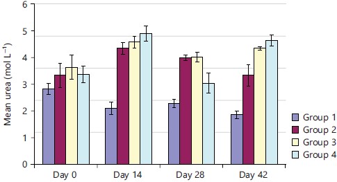
On day 0, there was no significant difference in the serum urea levels of the different groups compared to the control but as the administration continued, there were significant increases (p<0.05) in the urea levels on days 14, 28 and 42 when compared to the control group level as demonstrated in Fig. 7. This implied that continued administration of the extract caused an increase in the urea.
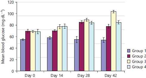
The blood glucose concentration of rats treated with ethanol extract in Fig. 8, significantly increased (p<0.05) on all the days compared to the control rats. The increase in blood glucose concentration was higher on days 28 and 42. On day 42, the glucose concentration in group 3 was highest compared to all the other groups and the control.
DISCUSSION
The ALT activity was observed more in group-2 (10 mg kg–1) and group-3 (50 mg kg–1) showing a significant increase (p<0.05) in the concentration of ALT compared to the control. There was a remarkable dose-dependent increase in AST activity from day 14 to day 42. This implied that the administration of ethanolic extract had an increased effect on AST concentration by doses and by the number of days. The extract caused a significant increase (p<0.05) in the concentration of ALP compared to the control implying that continuous administration of the ethanol extract caused an increase in the ALP concentration. There were slight increases in the level of total bilirubin among all groups from days 14, 28 to 42 when compared to the control group while group-4 (250 mg kg–1) on day 42 had a significantly higher concentration of total bilirubin compared to other groups. There was a gradual decline from day 14 in conjugated bilirubin concentration among the four groups. On days 28 and 42, groups 2 and 4 had no significant differences (p>0.05) in the creatinine concentration while in group 3, it was significantly higher than in the control group. There were significant increases (p<0.05) in the urea levels on days 14, 28 and 42 when compared to the control group level. The blood glucose concentration of rats treated with ethanol extract in significantly increased (p<0.05) on all the days compared to the control rats.
Since time immemorial, man has continued to depend on plant-based products to treat and manage diseases without understanding the mechanisms of action and adverse effects of some pharmaceutically bioactive principles12. Available information over the years revealed that improper intake of herbal products can cause severe liver and kidney injuries, acute and chronic abnormalities, anaemia, cirrhotic transformation and necrosis12. Hence, our investigation into the biochemical alterations associated with the oral administration of ethanol extracts of Nauclea latifolia roots in Wistar rats became necessary. The acute toxicity study of the ethanol extract in rats established an intraperitoneal LD50 greater than 4000 mg kg–1, thus, the ethanol extract could be generally regarded as safe. Phytochemical analysis of N. latifolia root extract showed the presence of sugars, saponins, terpenoids and flavonoids. This was in agreement with the work of Ugwoke and Obasi13. The constituents identified to belong to various classes of phytochemicals which are known to possess a wide range of pharmacological activities14. Thus, the anti-inflammatory, anti-oxidant, hypocholesterolemic and antibacterial activities reported for phthalates, aromatic hydrocarbons and fatty acids such as pentadecanoic acid, may suggest the rationale for the traditional uses of N. latifolia15,16. Furthermore, flavonoids have been widely described in the literature as vasodilator compounds. Saponins and flavonoids are also said to have other medicinal properties in animals17. These substances are known to have beneficial haematological and immunological properties. The antioxidant properties of flavonoids protect blood vessels' fragile capillaries against damage, however, high concentrations of the substances are toxic and may impair body metabolism18,19. Shigemori et al.20 reported the presence of indole alkaloids in the roots of N. latifolia, but no alkaloids were found in his study. This may probably be due to environmental variation in the areas where the plants were obtained (since the methods used for testing for alkaloids were the same).
Liver function tests (ALT, AST, ALP, total protein and bilirubin) provide information about the state of the liver by describing its functionality, cellular integrity and link with the biliary tract21. The results of the liver function tests in which serum enzyme activities of alkaline phosphatase, alanine aminotransferase, aspartate aminotransferase and the serum concentrations of conjugated and total bilirubin were measured seem to suggested mild damage to the liver hepatocytes of rats treated with ethanol extract of Nauclea latifolia root when compared to the control. Alanine Aminotransferases (ALT), a cytosolic enzyme whose activities increase as a result of cellular membrane damage, as well as Aspartate Aminotransferase (AST) are used for the detection of hepatocellular damage in animals22. The results of cytotoxicity analyses of Nauclea latifolia showed that it is non-hepatotoxic at 4000 mg kg–1 but, caused significant increases in the levels of ALT, AST and ALP. Besides the liver, AST is found in many other organs including the heart, kidney and brain. Thus a high level of AST does not always indicate that there is a liver problem. Alkaline Phosphatase (ALP) is a standard biomarker of biliary tract obstruction23. High levels of this enzyme in the serum may indicate cardiac infarction, muscle injury and hepatic necrosis24. Therefore, a high level of these enzymes observed in rats treated with Nauclea latifolia in this study, probably indicates that its prolonged and excessive use may be hepatotoxic. Transient increase of this enzyme may also be noticeable in all types of liver problems. Based on the results obtained, the extract of Nauclea latifolia may be slightly toxic. However, since the knowledge of the pharmacokinetics of a drug, apart from the dose, is also required in determining the degree of toxicity, toxicological studies on the extract are still being determined. More human and animal experimental studies are still needed to clarify this issue.
The extract caused a slight but not significant increase (p>0.05) in blood glucose concentration as administration of the extract continued on day 28 and 42. This can be explained simply by the high concentration of reducing sugars observed during the phytochemical analysis. However, the result of high glucose concentration observed in this work can be compared with that of Gidado et al.25, who observed a significant decrease (p<0.05) in blood glucose concentration in diabetic rats after administration of aqueous extracts of Nauclea latifolia leaves. This could be due to varying concentrations in the leaves of the plant and the length of administration of the extract, as this research work elapsed 42 days while Gidado et al.25, used 24 days. Also, Gidado et al.25 used the leaves while in this research work, the roots were used. Thus during the process of detoxification and deamination, some non-detoxified compounds in the ethanol extract may have destroyed the hepatocytes which are involved in detoxification than the kidney cells which are more involved in excretion.
CONCLUSION
The present study revealed elevated levels in biochemical parameters such as ALT, ALP, AST and serum albumin upon administration of crude extract of Nauclea latifolia. The reported bioactivities of the plant, including the results of biochemical parameters in this work, may largely be due to the presence of these secondary metabolites. Elevated levels in biochemical parameters could be a result of the effect of Nauclea latifolia extract on the liver and kidney functions respectively. Hence, it can be deduced that prolonged administration of Nauclea latifolia could lead to hepatic or renal toxicity and abnormalities.
SIGNIFICANCE STATEMENT
This study discovered an increase in biochemical parameters that indicate the possible toxicity of Nauclea latifolia. This study will help the researchers to uncover the critical areas of cytotoxicity of this plant that many researchers were not able to explore. Thus a new theory on its deleterious effect may be arrived at.
REFERENCES
- Cheikhyoussef, A., R.W. Summers and G. Kahaka, 2015. Qualitative and quantitative analysis of phytochemical compounds in Namibian Myrothamnus flabellifolius. Int. Sci. Technol. J. Namibia, 5: 71-83.
- Krishnaswamy, K., 2008. Traditional Indian spices and their health significance. Asia Pac. J. Clin. Nutr., 17: 265-268.
- Udobi, C.E., B.E. Umoh and E.G. Asuquo, 2017. Effects of the ethanol extract of the stem bark of Nauclea latifolia Smith [Rubiaceae] on certain biochemical and haematological indices of Swiss albino mice. Asian J. Med. Health, 6: 1-9.
- Balogun, M.E., E.E. Besong, D.C. Obu, M.S.U. Obu and S.F.A. Djobissie, 2016. Nauclea latifolia: A medicinal, economic and pharmacological review. Int. J. Plant Res., 6: 34-52.
- Udobre, A.S., E.I. Etim, J.A. Udobang, A.E. Udoh, A.E. Akpan and N.A. Ekpo, 2013. Antiplasmodial effect of the methanol extract of the stem bark of Nauclea Latifolia. Indo Am. J. Pharm. Res., 3: 6484-6489.
- Iyamah, P.C. and M. Idu, 2015. Ethnomedicinal survey of plants used in the treatment of malaria in Southern Nigeria. J. Ethnopharmacol., 173: 287-302.
- Abbah, J., S. Amos, B. Chindo, I. Ngazal and H.O. Vongtau et al., 2010. Pharmacological evidence favouring the use of Nauclea latifolia in malaria ethnopharmacy: Effects against nociception, inflammation and pyrexia in rats and mice. J. Ethnopharmacol., 127: 85-90.
- Agyare, C., V. Spiegler, H. Sarkodie, A. Asase, E. Liebau and A. Hensel, 2014. An ethnopharmacological survey and in vitro confirmation of the ethnopharmacological use of medicinal plants as anthelmintic remedies in the Ashanti Region, in the central part of Ghana. J. Ethnopharmacol., 158: 255-263.
- Tittikpina, N.K., C.E.C.C. Ejike, E. Castelluci-Estevam, M.J. Nasim and S. Griffin et al., 2016. Togo to go: Products and compounds derived from local plants for the treatment of diseases endemic in sub-Saharan Africa. Afr. J. Trad. Complement. Altern. Med., 13: 85-94.
- Ebbo, A.A., D. Sani, M.M. Suleiman, A. Ahmad and A.Z. Hassan, 2020. Acute and sub-chronic toxicity evaluation of the crude methanolic extract of Diospyros mespiliformis hochst ex a. Dc (ebenaceae) and its fractions. Toxicol. Rep., 7: 1138-1144.
- Harborne, J.B., 2008. Phytochemical Methods: A Guide to Modern Techniques of Plant Analysis. 3rd Edn., Springer, New Delhi, India, ISBN: 9780412572708, Pages: 302.
- Arunsi, U.O., G.C. Chinyere, K.O. Ngwogu, A.C. Ngwogu and O.C. Atasie et al., 2020. Evaluation of the biochemical, haematological and histopathological parameters of female Wistar rats fed with aqueous and ethanol extracts of Aspilia africana leaves. J. Herbmed Pharmacol., 9: 257-267.
- Ugwoke, C.E.C. and E.U. Obasi, 2019. Determination of the phytochemical potentials and antimicrobial properties of Nauclea latifolia leaves smith (Rubiaceae). World J. Med. Sci., 16: 86-90.
- Haudecoeur, R., M. Peuchmaur, B. Pérès, M. Rome, G.S. Taïwe, A. Boumendjel and B. Boucherle, 2018. Traditional uses, phytochemistry and pharmacological properties of African Nauclea species: A review. J. Ethnopharmacol., 212: 106-136.
- Kumar, P.P., S. Kumaravel and C. Lalitha, 2010. Screening of antioxidant activity, total phenolics and GC-MS study of Vitex negundo. Afr. J. Biochem. Res., 4: 191-195.
- Aparna, V., K.V. Dileep, P.K. Mandal, P. Karthe, C. Sadasivan and M. Haridas, 2012. Anti-inflammatory property of n-hexadecanoic acid: Structural evidence and kinetic assessment. Chem. Biol. Drug Des., 80: 434-439.
- Okwute, S.K. and C.S. Ohiakwu, 2021. Phytochemical and GCMS analysis of the leaves extracts of Nauclea Latifolia. J. Pharmacogn. Phytochem., 10: 116-120.
- Asekun, O.T. and B.A. Adeniyi, 2004. Antimicrobial and cytotoxic activities of the fruit essential oil of Xylopia aethiopica from Nigeria. Fitoterapia, 75: 368-370.
- Odukoya, O.A., S.I. Inya-Agha and O.O. Ilori, 2007. Immune boosting herbs: Lipid peroxidation in liver homogenate as index of activity. J. Pharmacol. Toxicol., 2: 190-195.
- Shigemori, H., T. Kagata, H. Ishiyama, F. Morah, A. Ohsaki, and J. Kobayashi, 2003. Naucleamides A-E, new monoterpene indole alkaloids from Nauclea latifolia. Chem. Pharm. Bull., 51: 58-61.
- Chanda, S., J. Parekh, Y. Vaghasiya, R. Dave, Y. Baravalia and R. Nair, 2015. Medicinal plants-from traditional use to toxicity assessment: A review. Int. J. Pharm. Sci. Res., 6: 2652-2670.
- Ozer, J., M. Ratner, M. Shaw, W. Bailey and S. Schomaker, 2008. The current state of serum biomarkers of hepatotoxicity. Toxicology, 245: 194-205.
- Thapa, B.R. and A. Walia, 2007. Liver function tests and their interpretation. Indian J. Pediatr., 74: 663-671.
- Debebe, M., M. Afework, E. Makonnen, A. Debella, B. Geleta and N. Gemeda, 2017. Evaluations of biochemical, hematological and histopathological parameters of subchronic administration of ethanol extract of Albizia gummifera seed in albino Wistar rat. J. Clin. Toxicol., 7: 1000337.
- Gidado, A., Ameh, D.A., S.E. Atawodi and S. Ibrahim, 2008. Hypoglycaemic activity of Nauclea latifolia Sm. (Rubiaceae) in experimental animals. Afr. J. Tradit. Complementary Altern. Med., 5: 201-208.
How to Cite this paper?
APA-7 Style
Udeh Sylvester,
M.C., Ogugua,
V.N., Ojochenemi,
E.Y., Parker,
E.J., Egba,
S., Anaduaka,
E., Ugwu,
O.P., Abu,
M.S., Iornenge,
T.J. (2023). Biochemical Effects of Nauclea latifolia Ethanolic Root Extract in Rats. Pharmacologia, 14(1), 9-20. https://doi.org/10.17311/pharma.2023.09.20
ACS Style
Udeh Sylvester,
M.C.; Ogugua,
V.N.; Ojochenemi,
E.Y.; Parker,
E.J.; Egba,
S.; Anaduaka,
E.; Ugwu,
O.P.; Abu,
M.S.; Iornenge,
T.J. Biochemical Effects of Nauclea latifolia Ethanolic Root Extract in Rats. Pharmacologia 2023, 14, 9-20. https://doi.org/10.17311/pharma.2023.09.20
AMA Style
Udeh Sylvester
MC, Ogugua
VN, Ojochenemi
EY, Parker
EJ, Egba
S, Anaduaka
E, Ugwu
OP, Abu
MS, Iornenge
TJ. Biochemical Effects of Nauclea latifolia Ethanolic Root Extract in Rats. Pharmacologia. 2023; 14(1): 9-20. https://doi.org/10.17311/pharma.2023.09.20
Chicago/Turabian Style
Udeh Sylvester, M., C., V. N. Ogugua, E. Y. Ojochenemi, E. J. Parker, S. Egba, E. Anaduaka, O. P. Ugwu, Michael Sunday Abu, and T. J. Iornenge.
2023. "Biochemical Effects of Nauclea latifolia Ethanolic Root Extract in Rats" Pharmacologia 14, no. 1: 9-20. https://doi.org/10.17311/pharma.2023.09.20

This work is licensed under a Creative Commons Attribution 4.0 International License.